|
|
 |
 The
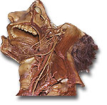
wax anatomical model production process involved four different
stages: The
wax anatomical model production process involved four different
stages:
1) preparation of an anatomical model by dissection of a cadaver;
2) construction of a plaster mould on the cadaver;
3) production of a model in colored wax;
4) finishing.
The first stage, essential to the production of a successful
model, was supervised by an expert dissector and involved the
sectioning of a cadaver to isolate the organs (or human body
parts) to be reproduced in wax.
In 1793, Dr. De Genettes, medical officer to the Napoleonic
armies in Italy, spent about a year at the Florentine Museo
della Specola (Observatory Museum) and left us a valuable record
of the wax modelling technique used by that particular School.
De Genettes wrote as follows:
"Most of the organs represented by colored waxes are
cast in plaster moulds formed directly from the natural organs.
The finishing touches are then added by a skilled sculptor
who works side-by-side with the actual cadaver. The sculptor
always works under the supervision of an anatomist, because
even the most excellent sculptors would be unable to reproduce
nature accurately without such guidance. Any organs that cannot
be cast directly are modelled in clay or wax by artists who
are extremely skilled in this type of work and copy from the
cadaver itself. A plaster impression is then cast on these
models. This technique is used particularly for statues of
whole figures.
When a plaster cast is to be made for an anatomical statue,
a sculptor is firstly commissioned to produce a full size
wax model. This is copied from life, nude and in the position
the anatomist finds that the organs or parts to be represented
are shown to the greatest effect. This first stage takes about
six months. When complete, separate models of individual organs
must be made once each has been dissected; and the entire
process must be carried out under constant supervision and
guidance."
Before proceeding to the second stage of
the work, the wax used to model the anatomical parts had to
be prepared. In the Florentine laboratory, from the end of
the Eighteenth century to the second half of the Nineteenth
century it was common practice to use a mixture obtained by
combining top quality beeswax with wax from other insects.
This Chinese wax has a high boiling point and is produced
by certain types of plant parasite. The resulting mixture
was poured into a big cauldron made out of tin-plated copper
and then left to melt over a very low heat. Lard (animal fat)
and sperm-oil (plant oil) were added to the mixture from time
to time as required.
The second stage involved the production
of a plaster cast which, when set, formed a negative impression
or mould, which was kept so that the same piece could be reproduced
over and over again if required.
The mould was generally cast directly from the anatomical
part, which was spread with a light layer of fat to protect
it from damage during the operation.
Now came the third and most delicate stage:
the production of the wax part. The success of this stage
depended upon the skill of the artist - and the artists employed
at the Specola Museum were unsurpassed in their proficiency.
Once the wax mixture had melted, a dye was added which had
already been dissolved in turpentine with the addition of
small quantities of powdered white lake. Natural dyes such
as madder lake, cinnabar and granulated white wax were used
to obtain the required muscle colors. Before the melted wax
was poured, the plaster cast was soaked in warm water and
its inner surface was spread with soft-soap. This made the
plaster less porous so that the model came away more easily.
The hot wax mixture was now poured into the mould. The operation
was carried out by pouring the wax in successive layers, each
at progressively lower temperatures.
Once the wax was cool, the mould was opened and the next stage
could begin.
The fourth and final stage involved cleaning
the resulting wax cast. This was carried out by scraping any
imperfections off the mould and polishing the model surface
with a soft brush soaked in oil of turpentine. The muscle
striations were then produced using heated iron implements
of various shapes and sizes. Blood vessels, nerves and lymphatic
vessels were then added: arteries, veins and nerves were produced
using wax-coated wire or thread. The finer vessels were painted
in using a very fine brush and colors were touched in as required.
The surface of the model was then covered in a coat of clear
varnish. This glaze added gloss to the model, protected it
from dust and muted the colors.
* taken from:
Various Authors,
Le Cere Anatomiche della Specola [Wax Anatomical models
of the Museo della Specola or Observatory Museum], Florence
1979
|
 |

|