|
|
 |
 |
 |
 Attempts
have been made to represent the whole human body, or body parts,
in wax (or more frequently terracotta or wood) since the very
earliest times. Archaeological excavation has revealed numerous
examples of this practice, part way between magic, medicine
and religion, in nearly all civilizations. In ancient Rome,
for example, the sick asked to be healed by appealing to the
gods (usually Aesculapius, god of medicine). Terracotta models
of organs were used to indicate the disease from which they
were suffering. Attempts
have been made to represent the whole human body, or body parts,
in wax (or more frequently terracotta or wood) since the very
earliest times. Archaeological excavation has revealed numerous
examples of this practice, part way between magic, medicine
and religion, in nearly all civilizations. In ancient Rome,
for example, the sick asked to be healed by appealing to the
gods (usually Aesculapius, god of medicine). Terracotta models
of organs were used to indicate the disease from which they
were suffering.
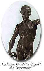
The first known anatomical representation of the human body
was a sculpture to which neither art nor science can lay claim.
This was the flayed man (so-called because the body is shown
without its skin) produced in 1600 by Lodovico Cardi (1559 -
1613), who was known as "Il Cigoli". The artist trained
in the Florentine school of Bronzino and soon developed a passion
for the discipline of artistic anatomy. He attended numerous
dissections of cadavers at the S. Maria Nuova Hospital in Florence.
The flayed man, a very fine statue in red wax measuring 61 cm
in height, displays all the fruits of his anatomical knowledge.
It can still be admired at the Bargello National Museum.
About a century later, another great artist, the Sicilian abbot
Gaetano Zumbo (1656-1701), who studied anatomy in Bologna, seat
of one of the most prestigious universities in the world, became
the very first to model anatomical models in different coloured
waxes.
The first wax model in different colours depicting human anatomy
that the Sicilian artist produced was a mans head: a stunning
masterpiece of anatomical realism. The wax head is kept in the
Museo della Specola (Observatory Museum) in Florence; original
home of the Cagliari wax anatomical models.
The heyday of wax anatomical modelling and medical and surgical
wax modelling in general did not really begin until the eighteenth
century, when the problem of training doctors, surgeons and
midwives (obstetricians) became increasingly pressing. Lecturers
found wax medical models were very useful in helping their young
students learn more about basic disciplines (anatomy, physiology,
surgical anatomy and zoology) and applied subjects (clinical
medicine and surgery, obstetrics etc.).The wax constructions
provided an enormously important teaching aid. They allowed
students to find out more about human body structures which
may be difficult to see during the dissection of a cadaver.
The models helped them to memorize the layout of difficult areas
and could be used to recreate the most common surgical operations
or the appearance of specific diseases.
A school of celebrated wax modellers began to form in Bologna
from the beginning of the Eighteenth century. These master craftsmen
were ultimately responsible for inspiring the Florentine artists
at the Museo della Specola (Observatory Museum). The Bolognese
Ercole Lelli (1702-1766), a painter, sculptor and architect
with a thirst for knowledge of human anatomy, decided to use
coloured wax models to teach students about the structure of
the human body without the need for dissection. Lelli produced
his anatomical preparations by molding rags and tow soaked in
wax and turpentine oil around real human skeletons. In 1742,
Lelli was commissioned by Pope Lambertini to produce a collection
of wax models for the University of Bologna Anatomy Laboratory.
Lelli was subsequently assisted by his contemporary the Bolognese
Giovanni Manzolini, (1700-1755), an artist (painter and sculptor)
and anatomist, who was aided in his work by his wife Anna Morandi(1716-1774). By the time her husband died, Anna Morandi had
acquired considerable skill as an anatomist and continued to
produce wax models. She became so proficient that she won fame
throughout Europe. Anna Morandi was probably the only woman
involved in wax anatomical modelling at that time and also taught
Human Anatomy at the University in Bologna.
The Florentine wax anatomical modelling
school was a direct offshoot of the Bolognese school and came
into being through the efforts of the Florentine surgeon Giuseppe
Galletti, professor of obstetrics in the main hospital of
S. Maria Nuova in Florence. Galletti went to Bologna to see
the work of Lelli and of the Manzolinis. He also saw obstetric
wax models produced by Giovanni Antonio Galli (1708-1782),
professor of obstetrics in Bologna. So captivated was he by
the beauty of these works of art that he endeavored to set
up a collection of wax obstetric models as soon as he returned
to Florence.
After many teething problems, Galletti eventually met the
modeller Giuseppe Ferrini and was able to create a series
of obstetric models in terracotta and wax, which were used
to demonstrate the various types of birth to students of medicine
and surgery and students studying in the school of Obstetrics.
In 1771, when the La Specola Natural History Museum was set
up (the most important museum of its kind in Florence and
all of Europe at its time), Ferrini began to work under the
guidance of Felice Fontana, who was founder of the famous
wax anatomical modelling workshop and employer of Clemente Susini, creator of the Cagliari wax models. Many other Italian
and foreign university and national museums possess wax anatomical
model collections produced in the Florentine school. Italian
museums with wax model collections include Florence, Bologna
and Cagliari, as already mentioned, and also Pavia, Pisa,
Modena and Trapani. Foreign museums with collections of wax
models include the Josephinum Military Academy of Health Museum
in Vienna, the British Museum in London, the Semmelweis Museum
in Budapest, the Anatomy Department Museum at the University
of Leyden in Holland, and others.
|
 |

|